
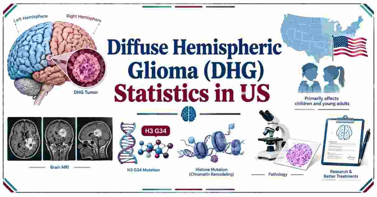
Diffuse hemispheric glioma, H3 G34-mutant (DHG, H3 G34), accounts for less than 1% of all gliomas diagnosed in adolescents and young adults, with a median age at diagnosis of 16 to 17 years across the largest published cohorts. Median overall survival ranges from approximately 17 to 22 months, with the largest combined dataset to date, drawn from 514 documented cases, reporting a median survival of 21 months.
Diffuse Hemispheric Glioma – Introduction
Diffuse hemispheric glioma, H3 G34-mutant is a rare and aggressive brain tumor formally recognized as a distinct diagnostic entity only since the World Health Organization’s 2021 classification of central nervous system tumors. Before that classification update, these tumors were often grouped together with pediatric glioblastoma, obscuring the distinct molecular biology and clinical behavior researchers have since identified. DHG, H3 G34 predominantly affects adolescents and young adults, arising in the cerebral hemispheres rather than the brainstem or midline structures affected by its better-known relative, diffuse midline glioma.
Because this tumor type has only been formally studied as its own category for a few years, published data still comes primarily from retrospective, multi-institutional case series rather than large prospective trials. This report compiles the most recently published statistics on DHG, H3 G34, from case volume and demographics to survival outcomes, prognostic factors, and molecular characteristics, drawing on the largest peer-reviewed cohorts published through 2026.
Interesting Facts About Diffuse Hemispheric Glioma in 2026
| Fact | Figure |
|---|---|
| Share of Gliomas in Adolescents/Young Adults | Less than 1% |
| Median Age at Diagnosis (Largest Combined Cohort) | 16 years (range: 1-66) |
| Median Overall Survival (Largest Combined Cohort) | 21 months |
| WHO Classification Grade | CNS WHO Grade 4 |
| Year Formally Defined as Distinct Entity | 2021 (WHO CNS5 classification) |
| Gross/Near-Total Resection Achieved (2026 Multi-Institutional Study) | 43% of patients |
| MGMT Promoter Methylation Rate | 75-82% across studies |
| Dominant Mutation Subtype | G34R (~93% of cases), vs. G34V (~6%) |
Data Source: Journal of Neuro-Oncology (Williams et al., 2025); American Association for Cancer Research Multi-Institutional Study (2026)
These figures describe a tumor type that, despite its rarity, has generated a meaningful and rapidly growing body of peer-reviewed clinical data since its formal recognition in 2021. The largest combined dataset assembled to date, published in the Journal of Neuro-Oncology, drew on 514 individually documented cases identified through both an institutional case series and a comprehensive literature review, with 257 of those cases including enough detail for individual-level survival analysis.
The 75% to 82% MGMT promoter methylation rate found across multiple independent cohorts is notably higher than the roughly 40% methylation rate typically seen in adult glioblastoma, a distinguishing molecular feature that has informed ongoing questions about whether temozolomide-based chemotherapy, which relies partly on MGMT status for its effectiveness, might play a meaningful role in treatment for at least some patients with this tumor type.
DHG H3 G34-Mutant Cases 2026 | Age & Demographic Data
MEDIAN AGE AT DIAGNOSIS — SELECTED DHG H3 G34 COHORTS
Pediatric-Only Cohort (≤18 years)       ██████████████████ 14 years
2026 Multi-Institutional Study (n=153)  ██████████████████████ 17 years
Largest Combined Dataset (n=514)        ████████████████████ 16 years
Molecular Landscape Study (n=114)       ████████████████████████ 22 years
Adult-Specific Cohort (n=17)            ██████████████████████████ 25 years
| Cohort | Sample Size | Median Age | Age Range |
|---|---|---|---|
| Pediatric-Only Cohort | 36 patients | 14 years | 8-18 years |
| 2026 Multi-Institutional Study | 153 patients | 17 years | 2-45 years |
| Largest Combined Literature Dataset | 514 patients | 16 years | 1-66 years |
| Molecular Landscape Study | 114 patients | 22 years | Not specified |
| Adult-Specific Cohort | 17 patients | 25 years | 19-33 years |
Data Source: PMC (American Association for Cancer Research, 2026); Journal of Neuro-Oncology (2025); Neuro-Oncology Advances
Across every major published cohort, DHG, H3 G34 consistently clusters around the adolescent and young adult age range, typically in the mid-to-late teens, though documented cases span an unusually wide range from early childhood through the sixth decade of life. The largest combined dataset, spanning 514 total cases, found a median age of 16 years with a range stretching from just 1 year old to 66 years old, illustrating that while the tumor is most characteristic of adolescence, it is not exclusively a pediatric disease.
Dedicated pediatric-only cohorts, restricted to patients 18 and younger, report a somewhat lower median age of 14 years, while cohorts specifically examining adult patients report considerably older median ages, around 22 to 25 years. This age distribution matters clinically, since several published analyses have specifically examined whether age at diagnosis independently predicts survival outcomes, a question covered in more detail in the prognostic factors section below.
DHG Survival Statistics 2026 | Overall & Progression-Free Survival
MEDIAN OVERALL SURVIVAL BY COHORT (months)
Pediatric-Only Cohort (≤18 years)                   ██████████████████████ 21.6
UK Adult, 13-Center Study (pediatric/AYA benchmark) █████████████████ 17.0
Largest Combined Dataset (n=514)                    ████████████████████ 21.0
Molecular Landscape Study (n=114)                   █████████████████████ 21.5
Midline Invasion Study (n=96)                       ██████████████ 14.4
Adult-Specific Cohort (n=17)                        ████████████ 12.4
| Cohort | Median Overall Survival | Median Progression-Free Survival |
|---|---|---|
| Pediatric-Only Cohort (≤18 years) | 21.6 months (1.8 years) | 8.4 months (0.7 years) |
| Pediatric/AYA Benchmark (UK Study Reference) | 17 months | ~10 months to progression |
| Largest Combined Dataset (514 cases) | 21 months | Not uniformly reported |
| Molecular Landscape Study (114 cases) | 21.5 months | Not uniformly reported |
| Midline Invasion Study (96 cases) | 14.4 months | Not uniformly reported |
| Adult-Specific Cohort (17 cases) | 12.4 months | Not uniformly reported |
Data Source: Multiple peer-reviewed cohorts, 2021-2026; see individual study citations throughout this report
Median overall survival for DHG, H3 G34 clusters in a fairly consistent range across most published cohorts, generally falling between 17 and 22 months, though individual study results vary based on cohort composition, treatment era, and follow-up completeness. The pediatric-specific cohort of 36 patients reported a median overall survival of 21.6 months, alongside a considerably shorter median progression-free survival of just 8.4 months, meaning many patients experience tumor progression well over a year before death, a pattern consistent with a disease that responds to initial treatment but recurs aggressively.
Notably, one adult-specific cohort study found shorter median survival, at 12.4 months, when comparing adult DHG, H3 G34 patients directly against other WHO grade 4 glioma types; that same study found no statistically significant survival difference between adult DHG, H3 G34 and either H3.3 K27M-mutant diffuse midline glioma or IDH-wildtype glioblastoma, but did find IDH-mutant glioblastoma patients survived significantly longer, a median of 50.5 months. For broader context on how DHG survival compares to the more common adult glioblastoma, see Glioblastoma Cancer Statistics in US.
DHG Prognostic Factors 2026 | What Predicts Better Outcomes
FACTORS LINKED TO IMPROVED SURVIVAL IN DHG, H3 G34
Gross/near-total resection (vs. subtotal/biopsy)     Statistically significant (p<0.001)
Right hemisphere tumor location                      Statistically significant (p<0.001)
G34R mutation (vs. G34V)                             Statistically significant (p=0.01)
Female sex                                           Positive factor (univariate)
MGMT promoter methylation                            Positive factor (univariate)
Absence of midline invasion                          Statistically significant
| Factor | Association With Survival |
|---|---|
| Gross/Near-Total Resection | Improved survival vs. subtotal resection or biopsy only (p<0.001) |
| Right Hemisphere Location | Longer survival vs. other locations (p<0.001) |
| G34R Mutation (vs. G34V) | Longer survival than G34V-mutant tumors (p=0.01) |
| Absence of Midline Invasion | Significantly better 12-month survival |
| Female Sex | Positive prognostic factor (univariate analysis) |
| Patient Age at Diagnosis | No significant independent effect in largest cohort |
Data Source: Journal of Neuro-Oncology (2025); Neuro-Oncology Advances (Molecular Landscape Study)
The extent of surgical resection consistently emerges as one of the strongest predictors of survival across published DHG, H3 G34 cohorts. The largest combined dataset found overall survival was significantly improved with gross total resection compared to subtotal resection or biopsy alone, a finding echoed in the 2026 multi-institutional AACR study, which specifically identified upfront gross or near-total resection, older age at diagnosis, and initial radiation therapy as factors associated with improved prognosis, even though that same study achieved gross or near-total resection in only 43% of patients, reflecting how often tumor location or extent limits safe surgical options.
Tumor location and molecular subtype also carry prognostic weight. Patients with tumors in the right hemisphere survived significantly longer than those with tumors elsewhere, and patients with the more common G34R mutation survived longer than those with the rarer G34V variant. A separate study focused specifically on midline invasion, defined as tumor spread into the contralateral hemisphere, brainstem, corpus callosum, thalamus, or basal ganglia, found this pattern present in 39.6% of the 96 patients studied and identified it as an independent predictor of significantly worse 12-month survival. Notably, one large analysis found no significant survival difference by patient age, MGMT promoter methylation status, or the presence of TP53, ATRX, or PDGFRA mutations, an unusual finding since age typically predicts outcomes strongly in most other high-grade gliomas.
DHG Molecular Features 2026 | Mutations & Genetic Markers
COMMON MOLECULAR FEATURES IN DHG, H3 G34 (% of tumors affected)
MGMT Promoter Methylation      ████████████████████████████████████ 75-82%
ATRX Loss/Negative Staining    ██████████████████████████████████████████ 93%
Olig2-Negative                 ████████████████████████████████████████████ 100%
TP53 Mutations                 ██████████████████████████████ Common
| Molecular Feature | Frequency |
|---|---|
| MGMT Promoter Methylation | 75-82% |
| ATRX Loss/Negative Immunostaining | 93% (adult cohort) |
| Olig2-Negative | 100% (adult cohort) |
| TP53 Mutations | Common, recurrent finding |
| CDKN2A/B Homozygous Deletions | Recurrent finding |
| PDGFRA, EGFR, CCND2, MYCN Amplifications | Recurrent findings |
| IDH Status | Always wildtype (IDH-negative) |
Data Source: Neuro-Oncology Advances, Molecular Landscape Study (114 patients)
DHG, H3 G34 carries a distinctive molecular signature that helps distinguish it both from IDH-mutant gliomas and from diffuse midline glioma, H3 K27-altered, its more widely known histone-mutant counterpart. Every documented DHG, H3 G34 tumor is IDH-wildtype, and molecular profiling studies consistently find loss of ATRX protein expression in the large majority of cases, alongside negative Olig2 staining, a marker typically present in other glioma subtypes. Recurrent genetic alterations include TP53 mutations, homozygous deletions of CDKN2A/B, and amplifications of PDGFRA, EGFR, CCND2, and MYCN, a pattern of alterations that molecular profiling researchers describe as helping refine the clinical and molecular landscape of this still relatively newly defined tumor type.
On imaging, DHG, H3 G34 tumors also present somewhat differently than typical glioblastoma, showing less avid contrast enhancement, less necrosis, and less surrounding edema on MRI, a pattern that one adult-focused study found led to an initial misdiagnosis as something other than a high-grade glioma in more than half of cases, underscoring the importance of molecular testing rather than imaging or histology alone for accurate diagnosis.
DHG in Adults vs. Children 2026 | How Outcomes Differ
DHG H3 G34 SURVIVAL — ADULT vs. PEDIATRIC/AYA BENCHMARK (months)
Pediatric/AYA Benchmark    █████████████████ 17.0
Adult-Specific Cohort      ████████████ 12.4
| Population | Median Overall Survival | Median Time to Progression |
|---|---|---|
| Pediatric/Adolescent Benchmark | 17 months | ~10 months |
| Adult-Specific Cohort (13 UK Centers) | Survival outcomes previously poorly characterized; under active study | Not fully established at time of study |
| Adult-Specific Comparison Cohort (17 patients) | 12.4 months | Not specified |
Data Source: PMC, 13-Center UK Retrospective Study; PMC, Adult DHG H3 G34 Comparison Study
Until relatively recently, survival outcomes for adult patients with DHG, H3 G34 remained poorly characterized, since most early research on this tumor type focused on the pediatric and adolescent population where it was first identified. A retrospective study spanning 13 UK hospital care centres was specifically designed to address this gap, noting explicitly that while pediatric and adolescent patients had an established median time to progression of roughly 10 months and median overall survival of 17 months, comparable adult-specific benchmarks had not been well established prior to that research effort.
Separate adult-focused comparison research, examining 17 adult patients, found a somewhat shorter median overall survival of 12.4 months, though the study’s authors cautioned this difference from pediatric/AYA benchmarks did not reach statistical significance given the small sample size involved. Median age at diagnosis in that adult cohort specifically was 25 years, within the broader 19-to-33-year range studied, reinforcing that even “adult” DHG, H3 G34 cases still cluster disproportionately in young adulthood rather than later in life. For a broader look at how pediatric brain tumor outcomes compare across the full range of childhood brain cancer types, see Brain Cancer Statistics in US.
DHG Treatment Approaches 2026 | Surgery, Radiation & MGMT Status
2026 MULTI-INSTITUTIONAL STUDY — TREATMENT PATTERNS (n=153)
Gross/Near-Total Resection Achieved      ████████████████████ 43%
Remaining Patients (subtotal/biopsy)     ████████████████████████████ 57%
| Treatment Factor | Detail |
|---|---|
| Gross/Near-Total Resection Rate | 43% of patients (2026 study, n=153) |
| Initial Radiation Therapy | Associated with improved prognosis |
| Temozolomide (TMZ) Response | Effectiveness in pediatric patients remains uncertain |
| MGMT Methylation Relevance | High rate (75-82%) raises open questions about chemosensitivity |
Data Source: American Association for Cancer Research, 2026 Multi-Institutional Study; PubMed Pediatric MGMT/TMZ Study
Current treatment for DHG, H3 G34 generally follows a similar framework to other high-grade gliomas: maximal safe surgical resection where feasible, followed by radiation therapy, with chemotherapy’s role remaining less clearly established. The 2026 multi-institutional study found initial radiation therapy was independently associated with improved outcomes alongside surgical resection extent, reinforcing radiation’s role as a standard component of treatment even in a disease with limited durable responses.
The role of temozolomide specifically in pediatric patients remains uncertain, according to a dedicated pediatric cohort study examining clinical, imaging, and molecular prognostic factors alongside MGMT expression and treatment response. That uncertainty persists despite the notably high rate of MGMT promoter methylation found across DHG, H3 G34 tumors generally, a molecular marker that typically predicts better response to temozolomide in other glioma types, suggesting that MGMT status alone may not translate into the same treatment benefit in this specific tumor type that it does in more common adult gliomas. Researchers reviewing the broader disease landscape have explicitly called for dedicated clinical trials and prospective patient registries, noting that current treatment approaches remain based primarily on extrapolation from other high-grade glioma protocols rather than DHG, H3 G34-specific evidence. For a broader picture of cancer statistics across the full range of malignancies affecting Americans, see Cancer Statistics in the US.
Frequently Asked Questions About Diffuse Hemispheric Glioma (DHG) in 2026
What is diffuse hemispheric glioma, H3 G34-mutant?
It is a rare, aggressive brain tumor formally recognized as a distinct entity in the World Health Organization’s 2021 classification of central nervous system tumors, predominantly affecting adolescents and young adults.
How common is DHG, H3 G34?
It accounts for less than 1% of all gliomas diagnosed in the adolescent and young adult population, making it a genuinely rare tumor type.
What is the median age at diagnosis for DHG, H3 G34?
Across the largest published cohorts, median age at diagnosis ranges from 14 to 17 years, though documented cases span from age 1 to 66.
What is the survival rate for DHG, H3 G34?
Median overall survival across most published cohorts ranges from approximately 17 to 22 months, with the largest combined dataset reporting a median of 21 months.
What factors improve survival in DHG, H3 G34?
Gross or near-total surgical resection, tumor location in the right hemisphere, the more common G34R mutation subtype (versus G34V), and initial radiation therapy are all associated with better outcomes.
How does DHG, H3 G34 differ from diffuse midline glioma?
DHG, H3 G34 arises in the cerebral hemispheres and carries a G34 histone mutation, while diffuse midline glioma typically involves a K27-altered histone mutation and arises in midline brain structures like the brainstem or thalamus.
Does age affect survival in DHG, H3 G34?
Findings are mixed. The largest combined literature dataset found no significant independent effect of age on survival, an unusual pattern compared to most other high-grade gliomas.
What is the MGMT methylation rate in DHG, H3 G34?
Between 75% and 82% of tumors show MGMT promoter methylation across published cohorts, notably higher than the roughly 40% rate typical in adult glioblastoma.
Is temozolomide effective for treating DHG, H3 G34?
Its effectiveness, particularly in pediatric patients, remains uncertain, and researchers have called for dedicated clinical trials to clarify its role.
How many total DHG, H3 G34 cases have been documented in medical literature?
The largest combined literature review identified 514 individual cases through January 2025, with 257 including sufficient detail for individual-level survival analysis.
Disclaimer: This research report is compiled from publicly available sources. While reasonable efforts have been made to ensure accuracy, no representation or warranty, express or implied, is given as to the completeness or reliability of the information. We accept no liability for any errors, omissions, losses, or damages of any kind arising from the use of this report.